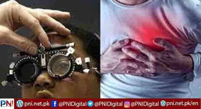
ISLAMABAD, JUNE 24 (ONLINE): Soon, retinal scans may be able to predict heart attacks. New research has found that decreased complexity in the blood vessels at the back of the retina in the human eye is an early biomarker for myocardial infarction.
“For decades, I’ve always lectured that the eye is not just the window to the soul, but the window to the brain and the window to the body as well,” said ophthalmologist Dr. Howard R. Krauss, speaking to Medical News Today about the new research.
“AI [artificial intelligence] plus ‘deep learning’ is proving that to be the case,” he said.
Cardiologist Dr. Rigved Tadwalkar, who was not involved in the research, told MNT that the findings were interesting.
“[A]lthough we have known that examination of retinal vasculature can produce insights on cardiovascular health, this study contributes to the evidence base that characteristics of the retinal vasculature can be used for individual risk prediction for myocardial infarction,” he said.
“This [study] representsanother tool in the toolbox to help determine who could potentially benefit from earlier preventative intervention [when it comes to heart attacks].”
— Dr. Rigved Tadwalkar
“The greatest appeal,” said Dr. Krauss, who was also not involved in the study, “is that the photography station may be remote to the clinician, and perhaps, someday, even accessible via a smartphone.”
The research was presented on June 12 at the European Society of Human Genetics.
Retinal scans and blood vessels
According to a press release, the project utilized data from the UK Biobank, which contains demographic, epidemiological, clinical, and genotyping data, as well as retinal images, for more than 500,000 individuals. Under demographic data, the data included individuals’ age, sex, smoking habits, systolic blood pressure, and body-mass index (BMI).
The researchers identified about 38,000 white-British participants, whose retinas had been scanned and who later had heart attacks. The biobank provided retinal fundus images and genotyping information for these individuals.
At the back of the retina, on either side where it connects to the optic nerve, are two large systems of blood vessels, or vasculature. In a healthy individual, each resembles a tree branch, with similarly complex fractal geometry.
For some people, however, this complexity is largely absent, and branching is greatly simplified.
In this research, an artificial intelligence (AI) and deep learning model revealed a connection between low retinal vascular complexity and coronary artery disease.
The power of AI
“The beauty of utilizing AI and deep learning is that as the database builds, one may learn of associations, and the predictive value of retinal evaluations, in ways which we may not even suspect today,” said Dr. Krauss.
Dr. Krauss added there are advantages of using AI and deep learning in such research.
“AI is capable of looking at a photograph and, with 97% accuracy, telling whether it’s a male or female. No ophthalmologist can look in the eye, or look at a photograph, and tell you if it’s male or female,” beyond guessing, he said.
The AI model was moderately successful when considering vascular density alone. However, Villaplana-Velasco described its accuracy as “significantly reduced when compared with a model that also included demographic data, and with established risk models.”
“Even when we just included age and sex to retinal vascular complexity, we found a significant improvement,” she said.
Specific genetic regions
A third factor improved the predictive power of the researchers’ model even further.
“Our genetic analysis showed,” said lead author and Ph.D. student Ana Villaplana-Velasco, “that four genetic regions associated with retinal vascular complexity have a role in MI-related biological processes.”
She said that her team was interested in further studying this link “by collaborating with other research groups focused on in-vitro experiments.”
“The findings make sense, in that a true association was seen between fractal dimension [complexity] and incident cardiovascular disease,” said Dr. Tadwalkar.
He noted that “the model also integrates [a polygenic] risk score, which can significantly improve precision on its own.”
Follow the PNI Facebook page for the latest news and updates.


